29+ brain and spinal cord drawing
Ascending and descending pathways Complete Table 1. Injuries of brain and cranial nerve with injuries of nerves and spinal cord at neck level.
Class Definition For Class 435 Chemistry Molecular Biology And Microbiology
T060 Sequelae of injuries of head.
. Item Preview remove-circle Share or Embed This Item. Keeping in mind you need no artistic skill whatsoever draw the brain and spinal cord. 2020-05-29 090319 Boxid IA1804205 Camera USB PTP Class Camera Collection_set.
Draw one cross section of the spinal cord from a slide and label all the areas you can. All spinal cord drawings ship within 48 hours and include a 30-day money-back guarantee. The remaining body was divided into anterior medial and posterior sections.
Use multiple colours to help distinguish each of the structures. You will learn more about the functions of these tracts Fig. The spinal cord extends from the medulla oblongate and is situated inside the vertebral canal.
The physician has the med tech draw blood for a CBC and to type and cross-match for blood. Choose your favorite spinal cord drawings from 29 available designs. Start studying Biology Honors Chapter 29.
An illustration of a magnifying glass. 13-5 Anatomy of the Spinal Cord Cylinder of nerve tissue within the vertebral canal thick as a finger vertebral column grows faster so in an adult the spinal cord only extends to L1 31 pairs of spinal nerves arise from cervical thoracic lumbar and sacral regions of the cord each cord segment gives rise to a pair of spinal nerves. The spinal cord is enclosed within but is shorter than the vertebral column.
In most adult mammals it occupies only the upper two-thirds of the vertebral canal as the growth of the bones. 299 202 to 446 Antigua and Barbuda. The regulation of homeostasis is governed by a specialized region in the brain.
A line drawing of the Internet Archive headquarters building façade. All tissues were fixed in 10 neutral buffered for-malin for histological examination. Brain -- Anatomy Spinal cord -- Anatomy Human dissection Central Nervous System -- anatomy histology Dissection.
The spinal cord is the central nervous system part that extends into the axial skeleton and provides the two-way traffic required to interact with our environment. A neurologist is a doctor who specializes in diagnosing and treating disorders of the brain and spinal cord. Harvey et al 2008 within FEAT and as recommended for use in PPI analyses Barton et al 2015.
For example afferent pain nociceptive signals can be inhibited yielding an anti-nociception that attenuates both spinally mediated withdrawal and brain-mediated indices of pain Fields 2000This provides a form of top-down processing that allows the organism to dynamically. T901-T902 T904-T905 T908-T909 If one or more of these codes was found in any of the diagnosis code fields in HDD NCHS Death or trauma registry data the record was determined to be a TBI. Traumatic brain injury TBI and spinal cord injury.
Then draw the meningeal layers Then draw the location of the skull and vertebral column that are there for protection. A direct nerve to muscle M-wave and the longer latency trans-spinally. Up to 15 cash back Find the perfect spinal cord drawing stock photo.
Drawing of brain and spinal cord At the end of this section you will be able to. An illustration of a magnifying glass. Physiological noise correction was conducted for the brain and spinal cord Brooks et al 2008.
Name the main regions of the adult brain Describing the relationship between the brain and the brain stem through diencephalon and from those areas into the spinal cord Recognize complex connections in the subcortical structure of the basalt nucleus Explaining the regulation of gray and. Where are the motor neuron cell bodies. The spinal cord is a long thin tubular bundle of nervous tissues that extends from the brain stem and continues down the center of the back to the pelvis.
Surgery of the brain and spinal cord based on personal experiences. Brain tissue and the anteriormost part of the spinal cord were removed and stored at 4 C to be processed later the same day for. Huge collection amazing choice 100 million high quality affordable.
Spinal cord grey matter can be divided in dorsal horn central grey and ventral hornThe dorsal horn comprises. The part of the nervous system that does not include the spinal cord and brain is known as the _____ nervous system. Neuron Structure Neurons are the functional cells of the nervous system.
Later development requires the contribution of neural. 28 in the Neurological Exam labLJXUH ³µ½ Major tracts of the spinal cord. Synaptic function in the spinal cord can be assessed using modification of the protocols widely used to measure motor and sensory nerve conduction velocity in rodents in which stimulation of the sciatic nerve via a needle electrode evokes two waveforms in muscles of the ipsilateral paw.
Where are the motor and sensory tracts. We use the 25th and 975th sorted values in the draw distributions as the upper and lower UIs for mean estimates and for percentage change whereby change was judged to be significant if the lower and upper UIs did not overlap zero. Interneurons- neurons in the brain and spinal cord that lie between the sensory and motor nerves and act as go-betweens or interpreters between the afferent and efferent nerves.
A line drawing of the Internet Archive headquarters building façade. Ascending tracts carry sensory information from the periphery to the brain. Learn vocabulary terms and more with flashcards games and other study tools.
Cardiac and respiratory phases were determined using a physiological noise model PNM part of FSL and slice specific regressors determined for the. It is highlighted in yellow in Figure 858. It is covered by the three membranes of the CNS ie the dura mater arachnoid and the innermost pia mater.
Research has shown that brain systems modulate the operation of spinal circuits. Based on the corresponding vertebrae level spinal cord is divided into the cervical C1-8 thoracic T1-12 lumbar L1-5 and sacral S1-5 segments. Figure 858 The spinal cord yellow runs from the bottom of the brain to the lower back.
The spinal cord is part of the central nervous system CNS which extends caudally and is protected by the bony structures of the vertebral column. During the exam your neurologist will test different functions of the nervous system. The types of tests will depend on your symptoms but most neurological exams include tests of some or all of the following.
During pregnancy early development of the spinal cord is influenced by the maternal dietary requirement for folate for closure of the neural tube. The cerebrum the diencephalon the brain stem and the cerebellum. A persons conscious experiences are based on neural activity in the brain.
R 11 for histology of nerve tissue and spinal cord See Chapter 12 for brain and spinal cord anatomy See Chapter 13 for cranial nerves and spinal nerves What you need to be able to do on the exam after completing this lab exercise. Drawing by yodiyim 1 94 Nervous control of muscle Clip Art by Blambs 11 343 3d spine Clipart by Eraxion 14 1041 Spinal cord Drawings by Eraxion 3 81 Spinal cord under pressure of bulging disc Stock Illustration by Alexmit 4 29 Control of defecation Stock Illustration by Blambs 2 301 Brain Anatomy - Sensorimotor area Stock. The spinal cord is a single structure whereas the adult brain is described in terms of four major regions.
In 2006 adult fish were obtained.

Free Mudroom Locker Plan With Storage Bench Pdf Blueprint White Laundry Rooms Room Layout Planner Bathroom Designs India
2

Via Giphy Massage Funny Massage Therapy Funny Massage Therapy Humor

Middle Earth Map Vector At Vectorified Com Collection Of Middle Earth Map Vector Free For Personal Use Middle Earth Map Map Painting Middle Earth

Tender Document Manualzz
Class Definition For Class 435 Chemistry Molecular Biology And Microbiology

Ray Logo Sun Ray Logo Handmade Logo Vintage Logo Boutique Etsy In 2022 Handmade Logo Rays Logo Shop Logo
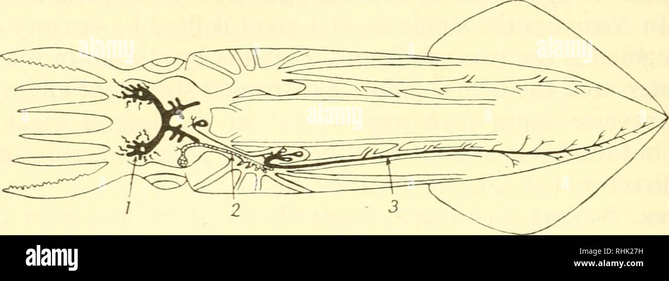
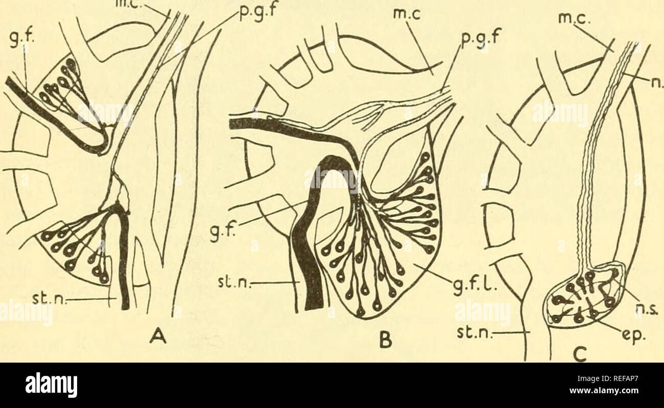
Stellate Ganglion High Resolution Stock Photography And Images Alamy
Class Definition For Class 435 Chemistry Molecular Biology And Microbiology
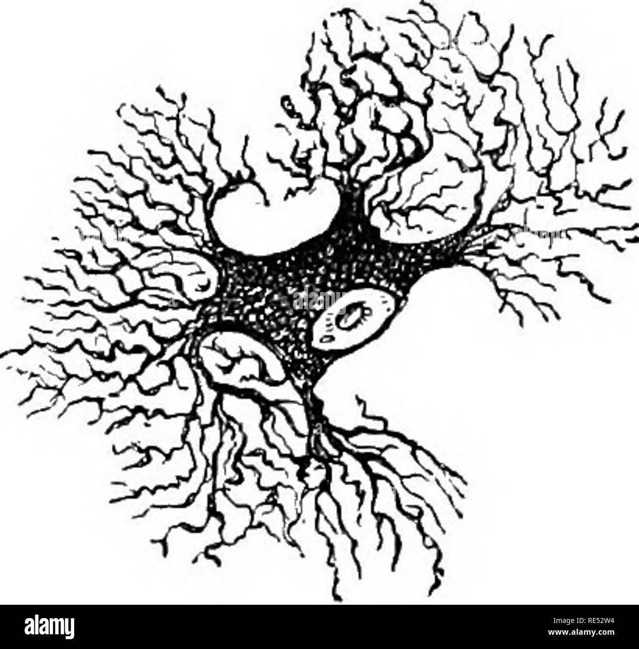
Stellate Ganglion High Resolution Stock Photography And Images Alamy
Class Definition For Class 435 Chemistry Molecular Biology And Microbiology
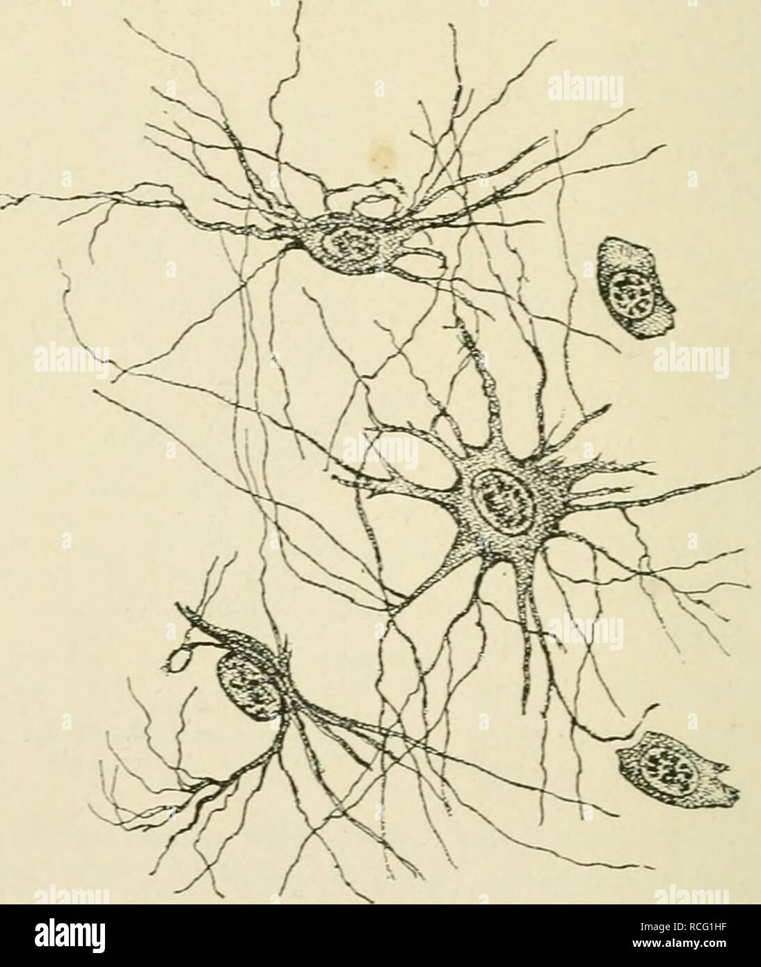
Stellate Ganglion High Resolution Stock Photography And Images Alamy
Stellate Ganglion High Resolution Stock Photography And Images Alamy

Factor Loadings And Factor Intercorrelations For The 3 Factor Model Download Scientific Diagram

Nurse Unicorn Medical Doctor Poster By Moon Ape 18 X 24 Doctor Medical Unicorn Doctor Stickers
29 Evidences For Macroevolution
2
![]()
Stellate Ganglion High Resolution Stock Photography And Images Alamy
Class Definition For Class 435 Chemistry Molecular Biology And Microbiology


Komentar
Posting Komentar